The discipline of military medicine necessitates an understanding of the neuroanatomy and neurophysiology of the motor unit. Nerve gases and their antidotes effect the third link, the neuromuscular junction, of the four-link chain which comprises the motor unit. Similarly, agents such as botulinum toxin with their bioterrorist potential also act at this site. Certain paralytic diseases for which military personnel receive vaccination (principally poliomyelitis) involve the first link in this chain 1. Envenomation by a range of poisonous creatures causes weakness or paralysis by damaging the second, third or fourth links of this basic structure.
The “motor unit” was the term coined by the Noble Laureate, Sir Charles Sherrington, in 1930, to define the functioning unit of motor neurones based in the spinal cord, medulla or mid-brain; their emergent axon; the neuromuscular junctions and the array of muscle fibres which contract following depolarisation2.
It is a delicious paradox that poisonous sea creatures which inadvertently can also cause human death and disability have been the agents by which neurophysiology and the discipline of toxinology has been advanced. In particular, the role of both venomous molluscs and toxic fish has been central to an understanding of the fundamental actions of the motor unit. Called by Sherrington “The Final Common Path”, an understanding of the function of the motor unit is central to the management of victims envenomed by a variety of land and sea creatures; and the prior protection or post-exposure clinical management of military personnel exposed to nerve gases.
Intoxications, envenomation’s or viral infections at any point along this four-link chain result in the subjective symptom of weakness; and the objective signs of hypotonia, flaccidity and paralysis. All who care for those presenting with acute weakness or paralysis needs raise a differential diagnosis which involves diseases along this four-link chain. The history of the advancement of knowledge – concerning the structure and function of the motor unit – has depended on the use of both molluscs and fish and their venoms, as experimental tools.
Differential Diagnosis – Acute Weakness and Paralysis
Any patient who presents with acute paralysis, weakness, hypotonia or flaccidity necessitates the raising of a differential diagnosis which includes the diseases that target one or more of the four links which comprise the motor unit chain3. Such diseases range from Sarin intoxication to snakebite, and from poliomyelitis to polymyositis4.
In 1669, the Dutch medical student, Jan Swammerdam, conducted the first experiments on nerve-muscle preparations, thus ushering in the modern era of experimentations which has provided an understanding of the motor unit. This in tum makes possible the interpretation of the symptoms and signs of envenomed and intoxicated patients today.
A significant milestone in the modem era of neuromuscular research was von Haller’s discovery, in Gottingen in 1757, that vertebrate muscles possess intrinsic irritability and contractility which are independent of any supplying nerve. Haller, a great polymath, was also a poet. One of his oft-quoted stanzas reads:
“Of Nature’s inmost heart no human mind can tell Happy, indeed, is he who knows its outer shell”
Claude Bernard (1813-1878} provided the final experimental proof that the contractility of muscles was due to electrical effects in the muscle fibres themselves. In 1856 he used curare to paralyse nerves in vertebrate nerve muscle preparations – and then demonstrated that the muscles could be stimulated to contract normally by direct electrical stimulation. These experiments formed the basis of our present-day understanding of post-envenomation rhabdomyolysis, muscular dystrophy and viral polymyositis.
Claude Bernard’s life illustrated the importance of both a broad perspective and focused application in the prosecution of biological research. He wrote:
“Put off your imagination, as you take off your overcoat when you enter the laboratory; but put it on again, as you do your overcoat when you leave the laboratory. Before the experiment and between whiles, let your imagination wrap you around….”.
It is this broad imagination, this wider love of nature, which is so important in research into envenomation and intoxication by poisonous creatures; and in an understanding of the pathophysiologic effects which result from exposure to agents such as anticholinesterase inhibitors and competitive antagonists which act at the motor end place of voluntary muscles.
There exists a paradox that some of the world’s most venomous creatures have provided the research tools by which the action of their venoms can not only be understood but by which the evolving knowledge of the motor unit has developed.
Molluscs
Members of the Phylum have been used extensively in the delineation of the structure and function of the motor unit. The giant squid, Loligo forbesii, has a single giant axon (Figure 1). It was used, from 1939, by the Nobel Laureates (1963}, Sir Alan Hodgkin, A.F. Huxley and the Australian, Sir John Eccles, to define the depolarisation chain in the axon; and the discovery of sodium and potassium channels in the membrane of excitable tissues8. In 1949, Sir Bernard Katz joined the team and introduced the voltage-clamp technique. This research tool facilitated the discovery of other ionic channels in both nerve and muscles.
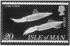
Figure 1: The Giant Squid, Loligo forbesii, whose single giant axon was the research tool used by Sir Alan Hodgkin, A.F. Huxley and Sir John Eccles in their discovery of ion channels and their role in the transmission of the depolarisation impulse along nerves. A 1994 Isle of postage stamp.
Another cephalopod, the Blue-ringed Octopus (Hapalochlaena sp.) was found to contain tetrodotoxin (TTX) (Figure 2). A low molecular weight substance which was found to block the sodium channel in neural tissues.

Figure 2: The Australian Blue-Ringed Octopus, Hapalochlaena maculosa, common in all eastern and northern Australian in-shore waters. Its saliva is a potent source of tetrodotoxin (TTX). An Australian 1984 postage stamp.
Tetrodotoxin was originally extracted from pufferfish (Figure 3). It occurs also in a Costa Rican frog and in poisonous newts (Family Salamandridae). Venoms from another family of molluscs, the cone shells (Canidae) have been used to define and study calcium channels in excitable tissues.
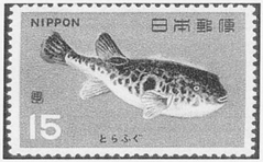
Figure 3: The Tiger Puffer Fish, Takifugu rubripes, an initial source of tetrodotoxin (TIX), used to block the sodium channel in excitable tissues. Tetrodotoxin is used as a basic research tool in the investigation of new venoms and toxins. From a 1966 Japanese postage stamp.
Omega-conotoxins are small polypeptides from Conus species, of which Conus geographus is the best known (Figure 4). Work currently being undertaken under the leadership of Professor Richard Lewis at the Centre for Drug Design at the University of Queensland has demonstrated that certain conotoxins act selectively on calcium channels in dorsal root ganglia, and thus possess the potential for novel pharmacological approaches to control severe and intractable pain.
The effects on acute short-term memory loss, consequent upon eating marine mussels contaminated with domoic acid, have been described following outbreaks of shellfish poisoning in Nova Scotia in 198710. The resultant intoxication syndrome, Amnesic Shellfish Poisoning, is distressing for the victims and perplexing for first aiders and doctors who manage these clinical effects. This toxin, derived from dinoflagellates of the genera Nitzchi and Pseudonitzchia, is concentrated in the tissues of filter-feeding bivalves (Class Pelecypoda) such as oysters, clams and mussels.
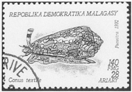
Figure 4: A 1992 stamp from the Malagasy Democratic Republic, showing a shell of the Family Canidae. The venom of cone shells contains, inter aHa, omega-conotoxins which have a selective calcium channel blocking effect on the neurones in the posterior root ganglia. Conus geographus has also caused human deaths by causing acute paralysis.
Fish
Certain coral reef and pelagic fish may concentrate a planktonic toxin, ciguatoxin, which is a cause of the dramatic human intoxication, ciguatera 11. Both the Pacific and Caribbean ciguatoxins cause their sensory, motor and autonomic effects12 by locking the sodium channel in the “open” position.
Historically, it was believed that the eating of terrestrial or marine snails of the family Turbinidae caused the enigmatic syndrome of ciguatera. The Spanish explorer in Cuba, Don Antonio Parra, in 1787 first described this intoxication. He used the local Creole word “cigua” to describe the marine or terrestrial gastropods which were believed to be its cause13. In 1774, when Captain James Cook sailed on H.M.S. Resolution to the south-west Pacific (near the island of New Caledonia) his crew became intoxicated with ciguatera following the eating of “Sea Bream”. His surgeon, the 28-year-old William Anderson RN. subsequently described the dramatic symptoms which afflicted both the crew and the ship’s dogs in that episode.
Because both tetrodotoxin and ciguatoxin the lock the sodium channel (in the “closed” and “open” positions respectively). They can be exploited in experimental studies of any new or unknown poison, toxin or venom – to test whether such new poisons act on the sodium channel.
Early research on the third link in Sherrington’s motor unit, the neuromuscular junction, was undertaken following studies of the acetylcholine receptor of certain fish. The electric organ of the electric eel, Electrophorus eleotricus and the electric ray (Torpedo sp.) contain the highest known concentrations of acetylcholine receptors9. This work was extended when it was found that alpha bungarotoxin, from the Banded Krait, Bungarus multioinctus, would bind selectively and irreversibly to the acetylcholine receptors of voluntary muscles. Radioactive bungarotoxin is thus used to facilitate the identification of and counting of acetylcholine receptors both for clinical studies of suspected myasthenia gravis and for neuromuscular research more generally.
Many molluscs, jellyfish, crabs, corals, ticks, scorpions, spiders and snakes possess poisons or venoms, the majority of whose actions remain unknown to science. Their further study holds great promise for development and better diagnostic tests; and certain novel therapies for human disease and envenomation.