Several rickettsial diseases have the potential to impact on military deployments. This article is a review of the information concerning the ecology, epidemiology and historical impact of rickettsial diseases on the Australian Defence Force (ADF) personnel in peace and in wartime. Probably the most widespread and important rickettsial disease is scrub typhus, while diseases such as tick typhus, murine typhus, epidemic typhus and Q fever have been of lesser importance.
Scrub typhus
Scrub typhus, an important rickettsial disease of humans, is widespread in central, eastern and southeast Asia, and the southeastern Pacific region.1 It is transmitted to humans by the bite of larval trombiculid mites, known as chiggers, infected with Orientia (formerly Rickettsia) tsutsugamushi, and causes significant morbidity and mortality. The epidemiology of scrub typhus is complex, involving the causative bacteria, vector chigger mites, vertebrate hosts that maintain the chigger mites, and humans, who by modifying the environment influences the ecology of maintenance hosts and is an occasional victim of the disease.2 The vectors of scrub typhus all belong to the genus Leptotrombidium. The main vector is L. deliense, which occurs in a wide area including southeast Asia, Pakistan and Australia, while L. akamushi, L. pallidum and L. scutellarae are found in Japan and Korea, and L. fletcheri and L. arenicola in Malaysia.1 A number of other genera of trombiculid mites have been found with O. tsutsugamushi in their tissues using direct fluorescent antibody tests, but their role as vectors of scrub typhus has not been confirmed.3, 4
Scrub typhus was a significant disease among allied troops operating in the south west Pacific region during World War II. In Papua New Guinea there were approximately 2,840 cases reported in the Australian Army between March 1942 up to December 1945 (39 months), of which 2,100 cases were reported before the end of 1943, with a case mortality rate of 9%.5,6 Studies on control measures against scrub typhus were facilitated by the isolation of the Karp strain of O. tsutsugamushi at Queensland Institute of Medical Research in 1943 from an American soldier (Karp) who had been wounded at Buna (on the north coast of Papua New Guinea) and evacuated to Brisbane.7 An Australian soldier, also wounded at Buna in December 1942 was photographed being taken to a field hospital by a New Guinea native (Photo 1), and although the soldier survived his battle injury, he unfortunately died from scrub typhus infection in February 1943.
The use of personal protection measures against mite vectors of scrub typhus were investigated in Australia and the USA in 1942-43. In 1942, American workers found that dimethylphthalate and other new mosquito repellents applied to military clothing were effective against chiggers.8 The Australian Army found dibutylphthalate was less toxic to mites than dimethylphthalate, but was more readily available in Australia and more resistant to washing and was introduced at the end of 1943 as “anti-mite” fluid. The introduction of dibutylphthalate as an antimite fluid was responsible for a 90% reduction in scrub typhus infections among Australian soldiers.9 This is one of the few examples where the use of personal protection measures resulted in a significant recorded reduction in vector borne disease.
During World War II, soldiers feared scrub typhus more than malaria, as there were drugs available for malaria treatment.6 Development of tetracycline antibiotics for the treatment of scrub typhus was begun during World War II, and gained impetus after the end of the war. The work of the US Army was significant, and resulted in the use of chloromycetin as a treatment for scrub typhus.10 A combination of successful antibiotic treatment and clothing treatment resulted in confidence that the disease could be managed successfully.
After World War II, the next time that scrub typhus was a problem for Australian soldiers was during the emergency in Malaya between 1955 and 1959. In 1956 only 4 cases of scrub typhus occurred in a battalion of soldiers, but 8 cases occurred in March 1957 after an operation in which the battalion traversed a mite infested area. The use of dibutylphthalate treatment of military clothing was the only preventative method of protection, and the use of “anti-mite” fluid was re-enforced. Despite this, 13 cases of scrub typhus were recorded between July and September 1957. A total of 27 cases were subsequently reported in a battalion group in Malaya between 1957 and 1959.11
Scrub typhus was also an important disease among soldiers during the Vietnam conflict. There were sporadic cases of scrub typhus among Australian soldiers in South Vietnam. Australian soldiers were issued with dibutylphthalate for protection against vector mites. In 1962, there was a small team of advisors in South Vietnam, and there were few cases of scrub typhus. In May 1965, a battalion group of soldiers was in the Ben Cat region and 2 cases of scrub typhus were observed in soldiers who did not use anti mite fluid. Australian medical staff thought that scrub typhus did not occur in the region, but following the two cases in 1965, they actively recommended the application of dibutylphthalate to clothing and boots to provide some protection against trombiculid mites. In September 1967, a soldier died from scrub typhus, having originally being diagnosed as having Japanese Encephalitis virus. This fatality highlighted the need for diagnostic services and reinforced the danger of scrub typhus to troops. A study in Phuc Thuy province, Vietnam, found 17% of 94 soldiers with fevers of unknown origin (FUO) were suffering from scrub typhus infection.11 The incidence of scrub typhus in French and American personnel during conflicts in Vietnam was also significant. Pages et al. 12 reported between 20-30% of FUO’s among French and American troops were due to scrub typhus.
Studies conducted in Malaysia showed that the antibiotic doxycycline could be used as a prophylactic agent against scrub typhus.13 Doxycycline is currently the first line malaria prophylaxis used by the Australian Defence Force14 and is effective against rickettsial diseases, as well as some sexually transmitted diseases. The use of doxycycline as a malaria prophylaxis provides protection against infections such as scrub typhus in personnel deployed to endemic regions in southeast Asia.
Scrub typhus is endemic in northern Australia, and is found in the wet tropics of Queensland and the Northern Territory. The disease was first recognized in the 1920’s, and was one of the most important health problems in the region. Cases of the disease occurred in areas of Queensland with high rainfall and humidity, and were associated with rainforest habitats on the wetter side of the 60 inch (1524mm) isohyet.15 The incidence of scrub typhus in Queensland has been low during the last 4 decades, with only a few published reports of cases.16,17 In the early 1990’s a new focus of disease was reported at Litchfield National Park in the Northern Territory. Five cases of scrub typhus occurred in a remote rainforest region, with 2 near fatal due to multisystem involvement.18 The mite vector, Leptotrombidium deliense, was found on rodents collected near the human cases.19 A subsequent death from scrub typhus was reported from a worker in Litchfield National Park in 1996.20 Cases have also been reported in Torres Strait islands 21 and a single case in Western Australia.22
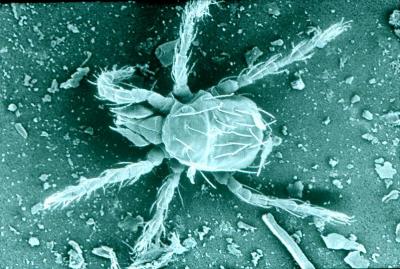
Personal protection against mites has also improved. In 1995, it was recommended that military uniforms be treated by dipping in a 0.6% water emulsion of permethrin (Perigen Defence®) to protect the wearer from mosquito-borne diseases malaria, dengue and other arboviruses. Permethrin treatment of clothing was shown to be effective against the scrub itch mite, Eutrombicula hirsti (Photo 2), during studies at Cowley Beach Training Area.23 There have been sporadic cases of scrub typhus among Australian soldiers in northern Queensland, and many of these have been reported from soldiers training at Cowley Beach.16, 17 The occurrence of cases in soldiers training at Cowley Beach (Photo 3) in 1996 prompted the recommendation that doxycycline prophylaxis be used by all ADF personnel training at Cowley Beach. This antibiotic is also recommended for prevention against leptospirosis.16
This disease is one of the few vector borne diseases that still causes a loss of manpower in Australia among ADF personnel, and members on operations or training in rainforest habitats will be adversely affected.24 Orientia tsutsugamushi occurs in a large area in southeast Asia and the south west Pacific region, and will continue to be a concern for deployed forces into the future.
Tick typhus The occurrence of tick-borne typhus is worldwide and is dependant on the species of tick present in a region. In Australia, two main species occur, Rickettsia australis, which is the causative agent of Queensland tick typhus,25 and R. honei, the cause of Flinders Island spotted fever. Queensland tick typhus was recognized as a new illness among Australian soldiers training on the Atherton Tableland during World War II.26,27 The disease was subsequently recognized in areas as far south as Victoria.28 The vector of R. australis is the Australian scrub tick, Ixodes holocyclus. This tick occurs along the eastern seaboard and ticks may remain attached to humans for several days while taking a large bloodmeal. R. australis causes a vesicular rash in humans, and is usually mild, although a single fatal case has been reported.29 Flinders island spotted fever was first described in 1991,30 and is caused by Rickettsia honei.31 The disease is characterised by fever, headache, myalgia, transient arthralgia, maculapapular rash and sometimes cough. The disease is also transmitted to humans by ticks.32 The finding of a new strain of spotted fever group rickettsia by Unsworth et al. 32 suggests that more genetically distinct strains may be discovered in the future. The new strain was designated the “marmionii” strain of R. honei, in honour of Australian physician and scientist, Barrie Marmion.32
Ticks are a nuisance for patrolling troops, but the occurrence of tick attachments in defence personnel is rarely reported in the scientific literature. Problems with tick attachment (Amblyomma triguttatum, kangaroo tick) to military personnel in Western Australia are an example where military training was adversely affected.33 The occurrence of R. australis in military personnel is minimal, although two cases were reported from soldiers training in the Cowley Beach Training area in 2005.17 Contact with ticks is enhanced by military activities, which include patrolling, resting and sleeping in forest habitats. These activities increase the likelihood of contact between military personnel and the tick vectors of typhus. The use of chemoprophylaxis for protection against Queensland tick typhus is not recommended, but the use of topical repellents and permethrin treated uniforms is thought to provide enhanced protection against tick attachment.34 Murine typhus (Rickettsia typhi).
Murine typhus has world wide distribution and is transmitted between rats by rat fleas (Xenopsylla cheopis). The mode of transmission of R. typhi to the vertebrate host was thought to be by infected flea faeces, and laboratory experiments showed transmission by the bites of fleas.35 The disease was first described by Dr Hone as occurring in Adelaide in Australia in 1922, In 1926 and 1960, cases of murine typhus were reported in Toowoomba, Queensland, associated with mouse plagues.36 Murine typhus has been subsequently reported in Mossman, Queensland 36 and Western Australia,37 suggesting that the disease is found Australia wide. Murine typhus caused infection in allied personnel during World War II and the Vietnam conflict. During World War II, the disease was not significant, but cases were identified in widely different areas, including north Queensland and Port Moresby, PNG.6 In Vietnam, murine typhus was rarely a serious illness, and the disease was treated with tetracycline. The disease was considered a threat before the first Gulf war when soldiers were deployed into endemic areas within Kuwait.12
Murine typhus is treated with a variety of antibiotics, with doxycycline being the most commonly used. This disease is rarely reported in Australia, with rural people most at risk, particularly during rat or mice plagues.
Epidemic typhus (Rickettsia prowazekii)
Historically, epidemic typhus caused by Rickettsia prowazekii has occurred in times of social unrest, war and famine. The disease is spread by the human body louse, Pediculus humanus. Lice become infected after ingesting blood containing rickettsia, which then enter the cells of the gut wall. Rickettsiae multiply until the cell bursts and rickettsiae are passed in the faeces of the louse. Transmission to humans occurs when louse faeces are scratched into the skin.38 Vigorous scratching of the area where lice have bitten allows rickettsiae to enter the body through the broken skin.
R. prowazekii caused major outbreaks of disease in many conflicts up to World War I. During World War II, due to advances in diagnostics, therapeutics, louse control methods and vaccine development, there were few cases of epidemic typhus. The disease can re-emerge due to the breakdown of social conditions as has occurred in some locations in Africa and the Middle East in the last 20 years.12 This disease does not occur in Australia, and environmental conditions in Australia limit the development and spread of body lice among people.28 The disease occurs in impoverished colder countries, often at high altitudes.
Q fever Q (Query) fever is a zoonosis with worldwide distribution. The disease was first described as a febrile illness in Brisbane abattoir workers.39 The causal agent was isolated from guinea pigs inoculated with patient blood by Dr Edward H. Derrick,40 and was originally called Rickettsia burnetii. The pathogen was re-named as Coxiella burnetii. The infection in humans is often non-specific and can be asymptomatic. Common symptoms include fever, fatigue, chills, myalgia, sweats and cough. The disease is transmitted to people in airborne droplets from infected cattle, sheep, rodents and cats. Although the organism is found in a number of tick species, human disease is acquired primarily through inhalation of aerosol droplets. Q fever is normally found in abattoir or dairy workers, but there have been recent incidental cases in military personnel, including British soldiers disposing of animal carcasses in the United Kingdom,41 Argentinean police working in Kosovo,42 and a small number of US soldiers in the middle east.43
Rickettsial agents as bioterrorist weapons The biological characteristics of rickettsiae and C. burnetii have allowed them to be weaponised for use as bioterrorism agents.44 The use of C. burnetii as a biological weapon would cause a disease similar to that of naturally occurring Q fever. The high infectivity rate means that only a small number of organisms would be needed to cause disease. The use of C. burnetii as an aerosol would be possible due to its resistance to desiccation, heat and persistence on wet and dry surfaces for a long time. There are limitations to the use of rickettsiae as biological weapons, including the need to produce highly purified, virulent, weapon quality rickettsiae that retain survival and virulence. The availability of counter-measures against bacteria may also limit their use as weapons.44 Despite these limitations, bioterrorism can instill fear into a society, devastate economies and cause diseases within the community.45
The serology of rickettsial diseases is the mainstay for diagnosis. This may involve collection of convalescent sera to confirm the diagnosis. All rickettsial diseases are treated with antibiotic therapy. Doxycycline is the drug of choice and chloramphenicol may be used as an alternative. The increased contact between soldiers and the vectors of rickettsial diseases, primarily ticks and mites, means that their risk of exposure to disease is increased. This is currently moderated by the use of personal protection measures against vectors and the use of antibiotics, which should provide good protection against disease. However, in some circumstances, these measures are not followed, cases of scrub typhus have occurred periodically, and so it is likely that rickettsial diseases will cause concern for military personnel in the future. The current frequency of deployments to warlike and humanitarian missions mean that rickettsial diseases will remain a threat to ADF personnel.